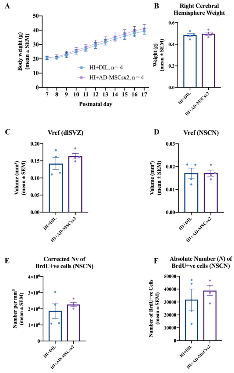
Figure 8.
Results from the experiment on cellular proliferation. Pups were exposed to either hypoxia-ischemia (HI) and diluent (HI + DIL) or HI and double treatment with adipose-derived-mesenchymal stem cells (HI + AD-MSCs×2). (A–F) Effect of delayed double treatment with either AD-MSCs or diluent on postnatal day (PN) 14/15 and PN16/17, after perinatal HI at PN7/8, on (A) the average body weight from PN7/8 to PN17, (B) the average right cerebral hemisphere brain weight at PN17/18, (C) the average volumetric data of the dorsolateral subventricular zone (dlSVZ), (D) the average volumetric data of the neural stem cell niche (NSCN) region within the dlSVZ, (E) the average neuronal density (Nv) of bromodeoyuridine (BrdU)-positive cells within the NSCN, and (F) the average absolute neuronal number (N) of BrdU-positive cells within the NSCN (HI + DIL: n = 4; HI + AD-MSCs×2: n = 4). All data are from the right cerebral hemisphere at PN17/18. The body weight data were statistically analyzed using used repeated measures ANOVA. The brain weight and stereological data were statistically analyzed using a paired two-tailed Student’s t-test. See also the text in the Results for more details on the statistical outcomes.

