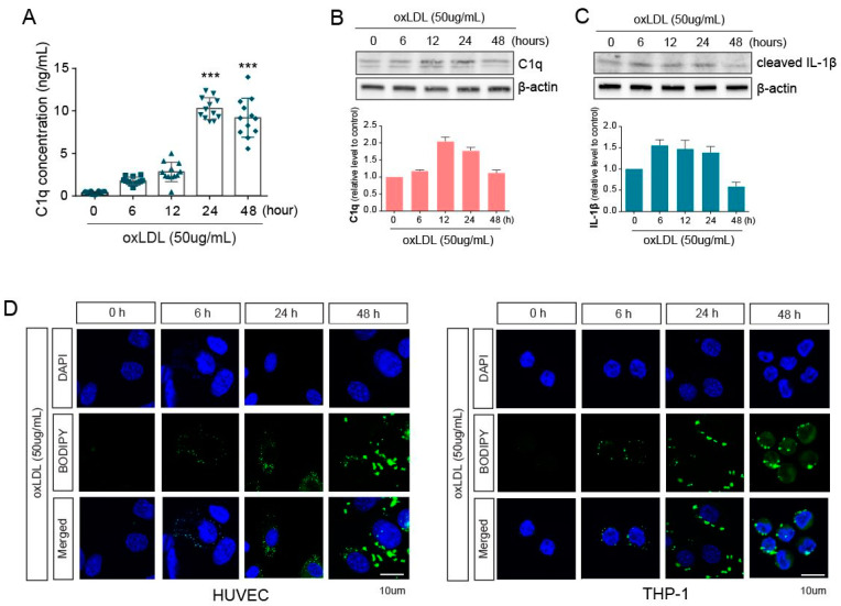
Figure 1.
Exposure of oxLDL on co-cultured macrophages and endothelial cells. (A) C1q concentration in the supernatant collected from co-culture media was measured using ELISA. (B) The protein levels of C1q in HUVECs were assessed by western blotting in whole-cell lysates at several time points after oxLDL exposure. (C) The protein levels of cleaved IL-1β were analyzed by western blotting in THP-1 whole-cell lysates at several time points after oxLDL exposure. (D) The accumulated lipids in HUVEC (left) and THP-1 (right) cells were stained with BODIPY and were detected by fluorescence microscopy. ***, p < 0.0001.

