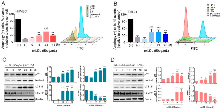
Figure 2.
Initiation of autophagy in co-cultured macrophages and endothelial cells upon oxLDL exposure. (A) After oxLDL treatment, autophagy in HUVECs was evaluated using the CYTO-ID kit and presented as relative values to the positive control. (B) Autophagy in THP-1 cells after oxLDL administration was evaluated using the CYTO-ID kit and presented as relative values to the positive control. (C) Western blot analysis in THP-1 cells was used in the evaluation of autophagy-related molecules. (D) Autophagy-related molecules were evaluated by western blotting in HUVECs. *, p < 0.05; **, p < 0.001, ***, p < 0.0001.

