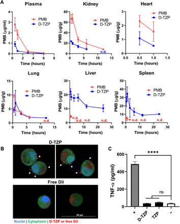
Fig. 5. D-TZP concentrates PMB in the liver and spleen and reduces macrophage response to LPS.

(A) Biodistribution of PMB in C57BL/6 mice after a single intravenous injection of D-TZP or PMB at a dose equivalent to 5 mg/kg. The data are shown as means ± SD (n = 3 mice per group per time point). n.d., not detected. (B) Confocal microscope z-section images of J774A.1 macrophages incubated with 1,1′-dioctadecyl-3,3,3′,3′-tetramethylindocarbocyanine perchlorate (DiI)–labeled D-TZP or free DiI for 4 hours. White arrowheads: D-TZP on cell surface. (C) Tumor necrosis factor–α (TNF-α) production from J774A.1 macrophages, preincubated with D-TZP or TZP at a concentration equivalent to 25 μg/ml PMB for 24 hours, thoroughly rinsed, and then challenged with LPS (10 ng/ml). +, LPS control; −, no challenge control. Statistical significance was accessed by Dunnett’s multiple comparisons test following ordinary one-way ANOVA (****P < 0.0001; ns, no significant difference). The data are shown as means ± SD (n = 3 independently and identically prepared batches).
