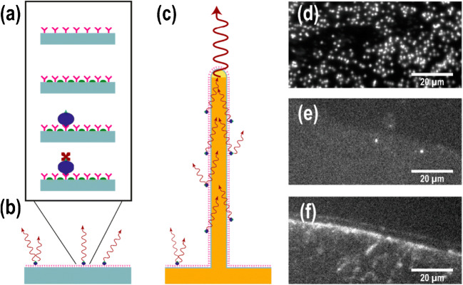
Fig. 1.
(a) A schematic of the single chain fragment variable (scFv) assay. First, the ScFv antibodies (shown in pink) were physiosorbed on the surface. The blocking agent (shown in green) was then utilized to reduce unspecific binding of biotinylated serum proteins (shown in blue). These serum proteins were targeted by fluorescently labelled streptavidin (shown in dark red). (b, c) A schematic of the assay on a flat substrate can be seen in (b) as well as a light-guiding nanowire substrate. (c) The supported waveguides to be reemitted at the tip were excited by the emission of fluorophores on the nanowire surface, which led to an enhanced signal at nanowires’ tip. (d–f) Epifluorescence images in top-view can be viewed of (d) a GaP nanowire substrate, (e) a silicon substrate, and (f) a MaxiSorp black polymer substrate, dotted with antibodies. The biotinylated serum concentration was diluted to 0.4% (which was the highest concentration used in this application). Individual nanowires can be seen in (d) as bright spots. The edge of the scFv spot is shown in (e,f), which shows the change in signal between specific and unspecific binding to the antibodies. Reproduced from Reference [27] with permission

