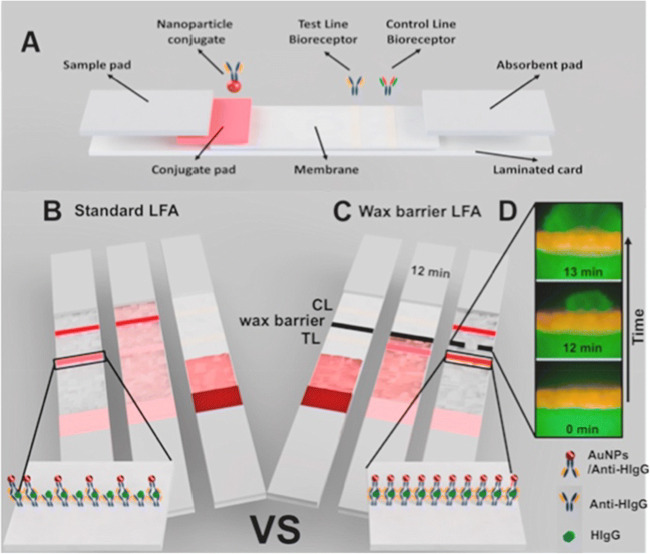
Fig. 5.
Strategy for the detection of proteins using wax barriers on top of a nitrocellulose membrane. (A) Schematic of different pads present in the LFA strip. (B) In the standard LFA, the flow constantly moved to the absorbent pad and the bio-recognition event occurred within seconds. Few labelled antibodies were captured in the test line (TL); therefore, the signal intensity was weak. (C) In the LFA modified with a wax barrier, the flow was temporarily stopped on the TL, resulting in increased time for the bio-recognition event. (D) Fluorescence microscope pictures (40×) of the wax barrier on the LFA strip. The wax barrier temporarily held the solution for 12 min. Reproduced with permission from reference [54]

