
Fig. 7.
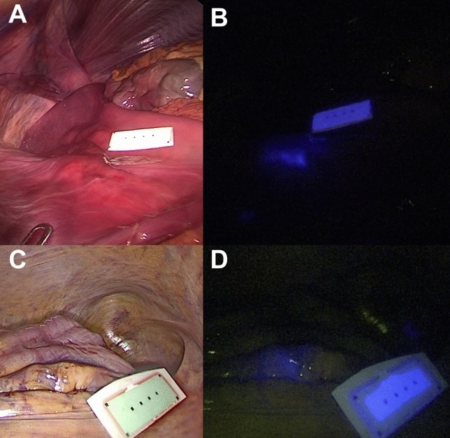
Fluorescent clip marking in the human anatomical specimen. Laparoscopic white light (left) and NIR imaging (right) mode visualization of the FOSC into the cadaver’s stomach (A, B) and sigmoid colon (C, D)
Official websites use .gov
A
.gov website belongs to an official
government organization in the United States.
Secure .gov websites use HTTPS
A lock (
) or https:// means you've safely
connected to the .gov website. Share sensitive
information only on official, secure websites.
Fluorescent clip marking in the human anatomical specimen. Laparoscopic white light (left) and NIR imaging (right) mode visualization of the FOSC into the cadaver’s stomach (A, B) and sigmoid colon (C, D)