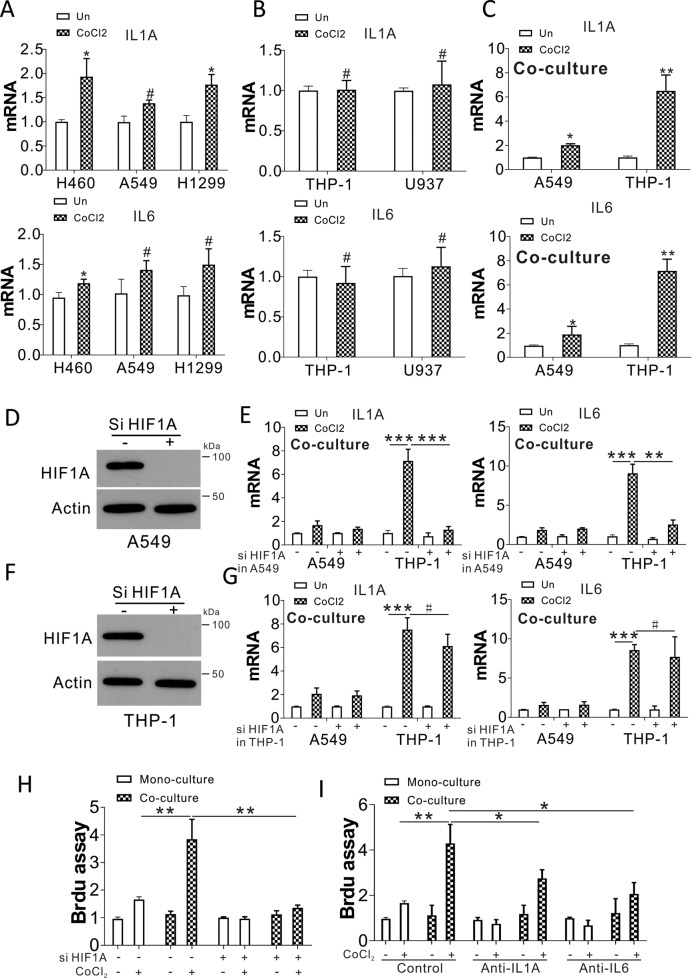
Fig. 2. Hypoxic stress in tumor cells mediates IL1A and IL6 expression in macrophages.
A IL1A and IL6 expression in H460, A549, and H1299 exposed to 100 µM CoCl2 for 24 h. B IL1A and IL6 expression in THP-1 and U937 cells exposed to 100 µM CoCl2 for 24 h. C IL1A and IL6 expression in A549 and THP-1 cells in the trans-well co-culture system with or without CoCl2 treatment in A549 cells. D HIF1α expression in A549 cells treated with HIF1α siRNA. E A549 cells transfected with control or HIF1α siRNA treated with CoCl2 for 1 d, and subsequently co-cultured with THP-1 cells for an additional day. IL1A and IL6 expression in A549 and THP-1 cells analyzed by RT-PCR. F HIF1α expression in THP-1 cells transfected with HIF1α siRNA. G A549 cells were treated with CoCl2 for 1 d and co-cultured with THP-1 cells with or without HIF1α siRNA transfection for an additional day. The expression of IL1A and IL6 in A549 and THP-1 cells was analyzed by RT-PCR. H The A549 cells transfected with control or HIF1α siRNA were treated with CoCl2 for 1 d, and co-cultured with THP-1 cells for an additional day. A549 proliferation was studied with a BrdU assay. I A549 cells were exposed to CoCl2 for 1 d, and co-cultured with THP-1 cells with or without anti-IL1A (MABp1, 10 ng/ml) or anti-IL6 (Sirukumab, 20 ng/ml) treatment for a further 24 h. A549 proliferation was studied using a BrdU assay. # p > 0.05; * p < 0.05; ** p < 0.01; *** p < 0.001.

