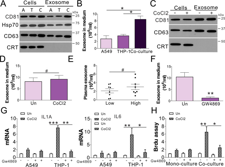
Fig. 3. Exosomes secreted by lung cancer cells trigger macrophages and promote inflammation.
A The exosomal positive markers CD81, CD63, and Hsp90 were observed in cells and media of A549, THP-1 mono-, and co-culture. Calreticulin expression served as a negative marker. A: A548 cells; T: THP-1 cells; C: co-culture. B Exosome levels in the cell culture medium of A549, THP-1 mono-, and co-culture. C A549 cells were treated with CoCl2 for 1 d, and co-cultured with THP-1 cells for another day. CD63, CD81, and CRT expression in co-cultured cells and media were studied through western blotting. D Exosome levels in the cell culture medium in (C). E Exosome levels in the plasma of patients with lung cancer with low and high HIF1α expression (n = 8 in each group). F The exosome level in the medium of A549 co-cultured with THP-1 exposed to 10 µM GW4869. G A549 co-cultured with THP-1 exposed to 10 µM GW4869. IL1A and IL6 expression was studied through RT-PCR. H A549 cells were treated with CoCl2 for 1 d, and co-cultured with THP-1 cells with or without 10 µM GW4869 for a further 24 h. The growth of A549 was analyzed by BrdU assay. # p > 0.05; * p < 0.05; ** p < 0.01; *** p < 0.001.

