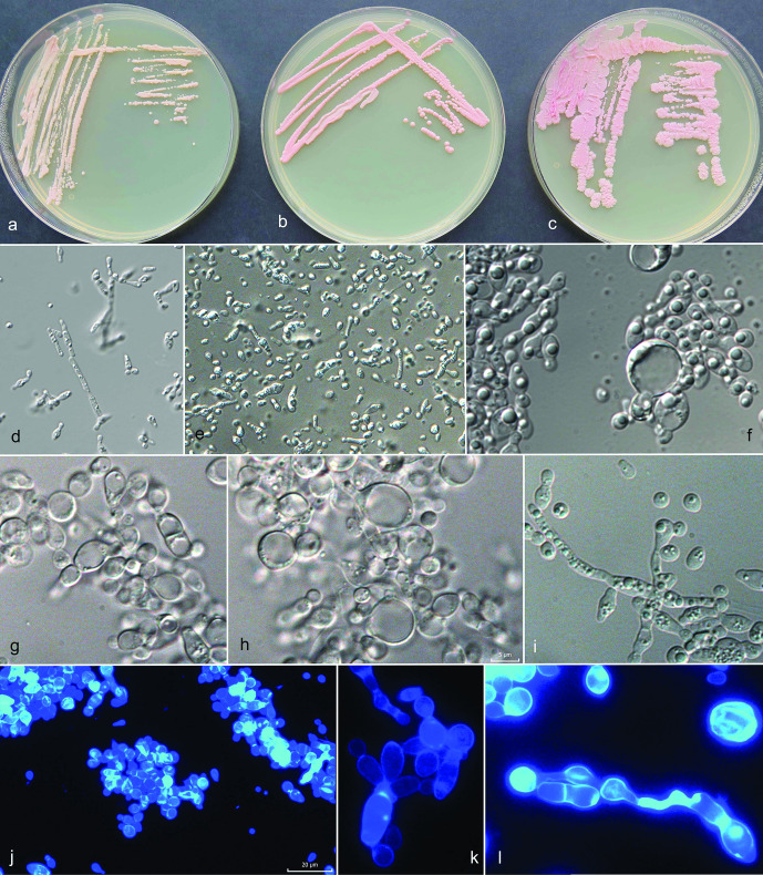
Fig. 3.
Morphology of Camptobasidium arcticum. (a–c) Cultures of Camptobasidium arcticum on PDA in 9 cm Petri dishes after 2 months of incubation at 15 °C: (a) EXF-12713T, (b) EXF-12689, (c) EXF-13086. Micromorphology of cells in water: (d) EXF-12689 on PDA after 14 days, (e) after 2 months, (f) EXF-12713T on PDA after 14 days, (g, h) on OA after 2 months, (i) EXF-12689 on OA forming pseudohyphae, (j–l) EXF-12713T on OA stained with calcofluor white. Scale bar indicated on fig. (j) (20 µm) is valid also for figures (d) and (e) — ×400 magnification; scale bar indicated on fig. (h) (5 µm) is valid also for figures (f), (g), (i), (k), (l) – ×1000 magnification.

