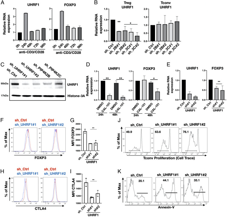
Fig. 5.
PIP4K depletion decreases UHRF1 levels in Tregs altering immunosuppressive capacity and cell viability. (A) Treg cells were TCR stimulated for the times indicated, and the expression of UHRF1 and FOXP3 was measured by qRT-PCR. (B) Expression levels of UHRF1 in Tregs (Left) and Tconv (Right) cells upon PIP4K silencing as indicated was analyzed using qRT-PCR. The empty pLKO_1 was used as control (sh_Ctrl) (n = 3). (C) Western blotting analysis of the expression levels of UHRF1 in Treg cells depleted of PIP4K or UHRF1 as indicated compared to control cells (sh_Ctrl). (D) RT-qPCR analysis of mRNA levels of UHRF1 (Left) or FOXP3 (Right) in Tregs treated with p110δ inhibitor (CAL-101, 10 μM) for 24 or 48 h (n = 3). DMSO was used as vehicle control. (E) UHRF1 was silenced in Tregs (sh_UHRF1), and the expression of UHRF1 (Left) and of FOXP3 (Right) were measured using qRT-PCR (n = 3). (F–I) UHRF1 was silenced, and protein levels of FOXP3 (F) or CTLA4 (H) were measured by FACS. (G and I) Quantitation of mean fluorescence intensity (MFI) values of FOXP3 and CTLA4 (n = 3). (J) UHRF1 was silenced in Tregs, and their immunosuppressive activity toward T conv cells was assessed (representative of n = 3). (K) UHRF1 was silenced in Tregs, and apoptosis was assessed by Annexin V staining (representative of n = 3). The charts represent means with SD. Statistical analyses were performed using unpaired Student’s t test, *P < 0.05, **P < 0.01. The number of replicates (n) refers to individual healthy donors.

