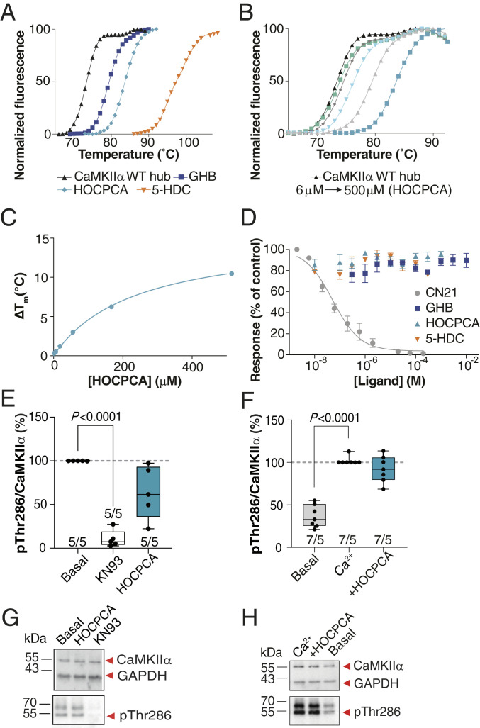
Fig. 3.
GHB analogs stabilize the hub but fail to affect CaMKII enzymatic activity under basal, nonpathological conditions. (A) Right-shifted thermal shift assay melting curves of CaMKIIα WT hub upon binding of GHB, HOCPCA and 5-HDC, (B) HOCPCA concentration dependence, and (C) saturation isotherm; representative data (see also SI Appendix, Fig. S5 D and E). (D) GHB analogs do not affect syntide-II phosphorylation by CaMKIIα, CN21 as positive control, pooled data (n = 3, means ± SEM). (E and F) CaMKIIα Thr286 autophoshorylation quantified by Western blot in cultured cortical neurons (days in vitro [DIV] 18 to 20). No effect of HOCPCA under (E) basal and (F) Ca2+-stimulated conditions. Shown is quantification of mean band intensities of Ca2+-stimulated pThr286 levels normalized to total CaMKIIα expression with 50 to 100 μM Ca2+ alone or together with HOCPCA (3 mM) for 1 h. (G and H) Respective representative Western blots. GAPDH was used as loading control. (E and F) Number in bar diagrams indicates number of experiments/individual cultures. Box plots (boxes, 25 to 75%; whiskers, minimum and maximum; lines, median) (one-way ANOVA, post hoc Dunnett’s test).

