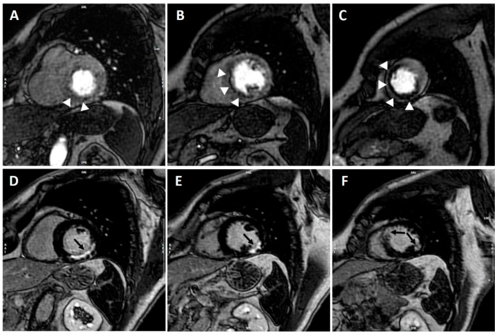
Figure 1.
CMR adenosine-stress perfusion in a 60-year-old patient with previous history of myocardial infarction. Short axis stress perfusion images at the basal (A), mid-ventricular (B), and apical (C) level. There is evidence of extensive inducible perfusion defect of the basal infero-septum and inferior wall, mid-cavity septum and apical anterior, and inferior and septal walls (white arrow-heads). Corresponding LGE images (D–F) show almost transmural LGE involving the basal inferior and infero-lateral walls, the mid-cavity infero-lateral wall, and the apical antero-septum and infero-lateral wall (black arrows). CMR findings were consistent with transmural myocardial infarction in the left circumflex territory with extensive inducible ischemia in the LAD territory; coronary angiography demonstrated critical proximal LAD stenosis.

