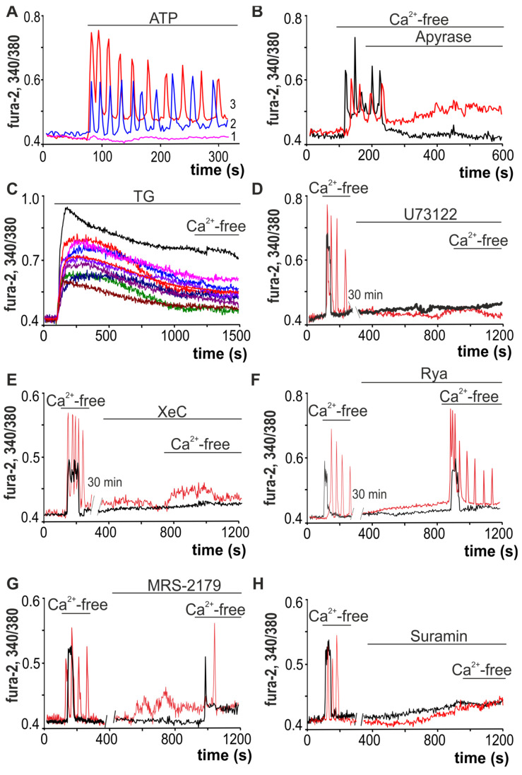
Figure 5.
Mechanisms underlying the Ca2+ responses of white adipocytes to decreases in external Ca2+. (A) Application of 10 µM ATP induces the generation of Ca2+ oscillations without changing the baseline [Ca2+]i level in 22 ± 16% of adipocytes (2) and with an increased baseline of [Ca2+]i level in 47 ± 11% of adipocytes (3), while in 31 ± 11% of adipocytes Ca2+ signals are absent. (B) Application of apyrase (apyrase, 35 units/mL), an enzyme that hydrolyzes ATP, against the background of Ca2+-free medium-induced Ca2+ oscillations, leads to their rapid and complete inhibition. (C) Ca2+-free medium-induced Ca2+ rises are prevented by discharge of the TG-dependent stores with 10 µM thapsigargin (TG). (D–F) Ca2+ signals due to the application of the Ca2+-free medium are completely suppressed by PLC (U73122, 10 µM, (D)) and IP3R (XeC, Xestospongin C, 1 µM, (E)) inhibitors and do not depend on RyR inhibition (Rya, Ryanodine, 10 µM, (F)). (G) Ca2+-free medium-induced Ca2+ oscillations of white adipocytes are suppressed by the P2Y1-receptor antagonist—MRS-2179 (30 µM)—but the Ca2+ signals have transient shapes. (H) Ca2+ signals of the white adipocytes after application of Ca2+-free medium are completely suppressed in the presence of suramin, an uncoupler of G-proteins and an antagonist of the P2X and P2Y receptors.

