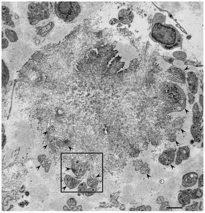
Figure 1.
Representative electron microscopy photograph of Schwann cells near a mass of amyloid fibrils. A cross-section of the sural nerve biopsy specimen from a patient with ATTRv amyloidosis. Subunits of Schwann cells indicated by arrowheads are located in the periphery of a mass of amyloid fibrils [28]. A high-powered view in the box is shown in Figure 2. Uranyl acetate and lead citrate stain. Scale bar = 2 μm.

