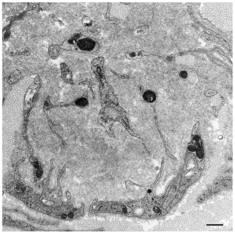
Figure 3.
Subunits of Schwann cells surrounded by amyloid fibrils in AL amyloidosis. A cross-section of the sural nerve biopsy specimen. Schwann cells become atrophic and their basement and cytoplasmic membranes are indistinct [21]. Uranyl acetate and lead citrate stain. Scale bar = 0.5 μm.

