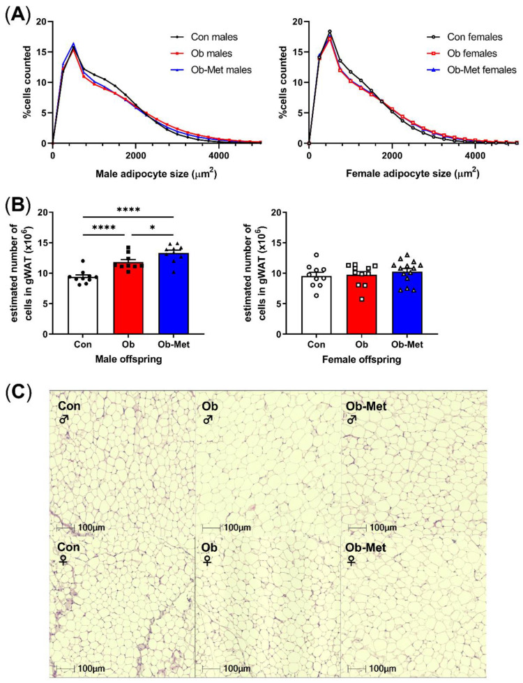
Figure 2.
Cellularity of 8-week-old gonadal adipocytes. (A) Adipocyte size distribution in male (left) and female (right) 8-week-old offspring. (B) Estimated adipocyte number in the collected gonadal white adipose tissue (WAT) depot. * p < 0.05, **** p < 0.0001 using one-way ANOVA with Tukey’s multiple comparison test. (C) Representative images of H&E-stained sections used for cell size analysis. Con (circles, solid line) = offspring of control-fed dams, Ob (squares, dashed line) = offspring of obese dams, Ob-Met (triangles, dotted and dashed line) = offspring of obese metformin-treated dams. Closed symbols denote male offspring; open symbols denote female offspring. n = 9–14 independent litters per group. Outliers were excluded from (B); male Con (Grubb’s method, outlier excluded value 6.28 × 10⁶).

