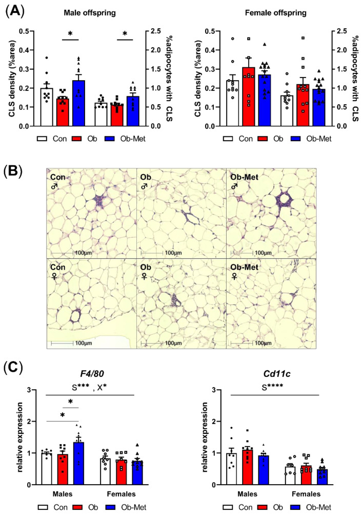
Figure 3.
Inflammation in gonadal white adipose tissue. (A) Presence of crown-like structures (CLS) in gonadal white adipose tissue (WAT) of 8-week-old male (left) and female (right) offspring, expressed as percentage area of the WAT tissue and as the number of CLS divided by the number of adipocytes in the section (n = 10–15 independent litters per group). * p < 0.05 using one-way ANOVA with Tukey’s multiple comparison test. (B) Representative images of H&E-stained sections used for CLS analysis. (C) mRNA expression of macrophage markers in gonadal WAT relative to the expression of housekeeper gene Ppia and the male Con group (n = 7–10 independent litters per group). * p < 0.05, *** p < 0.001, **** p < 0.0001 for the main effect of sex (S), interaction between sex and the maternal environment (X) or Tukey’s multiple comparison using two-way ANOVA. Outliers were excluded from (A) CLS density; female Con (Grubb’s method, outlier excluded value 0.46%), and (A) %adipocytes with CLS; male Con (Grubb’s method, outlier excluded value 1.19%). From (C) F4/80, two data points were excluded due to pipetting errors (male Con and female Ob). From (C) Cd11c, one data point was excluded due to pipetting errors (female Con).

