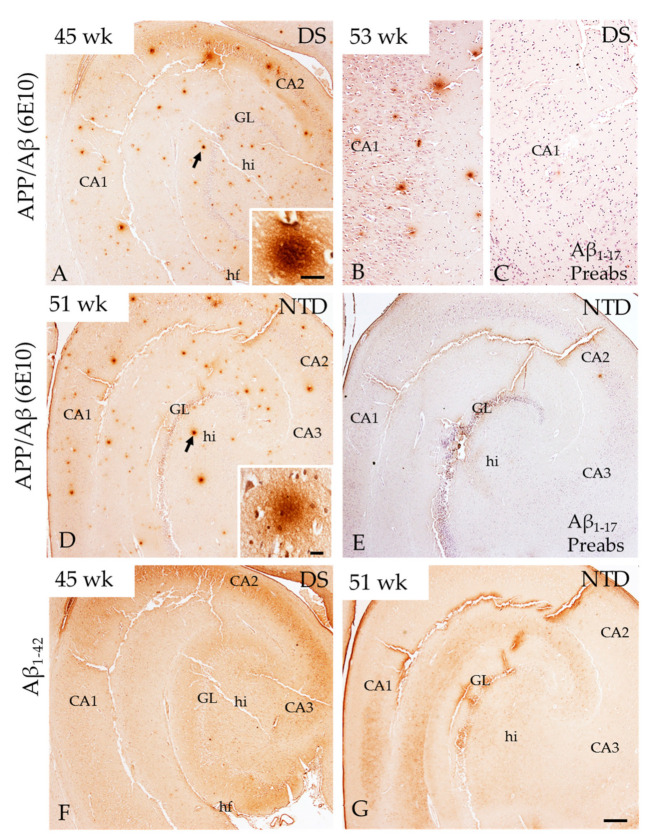
Figure 4.
Images of APP/Aβ (6E10) (A,B,D) and Aβ1–42 (F,G) immunopositive profiles in the hippocampus at postnatal week 45 and 53 in DS, and at week 51 in a NTD subject. High magnification images (insets) of APP/Aβ-ir profiles (arrows) in DS (A) and NTD (D). Note the random distribution of APP/Aβ-ir and absence of Aβ1–42-ir positive deposits in the hippocampus in both DS (A) and NTD (D). Additionally, absence of APP/Aβ-ir was noted in the hippocampus at postnatal week 53 (C) compared to antibody staining in panel B and similarly at 51 week in panel (E) compared to (D) in NTD (E) after antibody preabsorption with the Aβ1–17 peptide. Sections in (B,C,E) were counterstained with hematoxylin. Abbreviations: CA1 = hippocampal subfield CA1, CA2 = hippocampal subfield CA2, CA3 = hippocampal subfield CA3, fi = hippocampal fimbria, GL = granule cell layer, hi = hilus, hf = hippocampal fissure, Aβ1–17 Preabs= preabsorption with Aβ1–17 peptide, Sub = subiculum. Scale bar in (G) = 500 µm applies to (A,D–F) and in (B) and (C) = 100 µm; insets: (A) = 20 µm and (B) = 25 µm.

