
Figure 7.
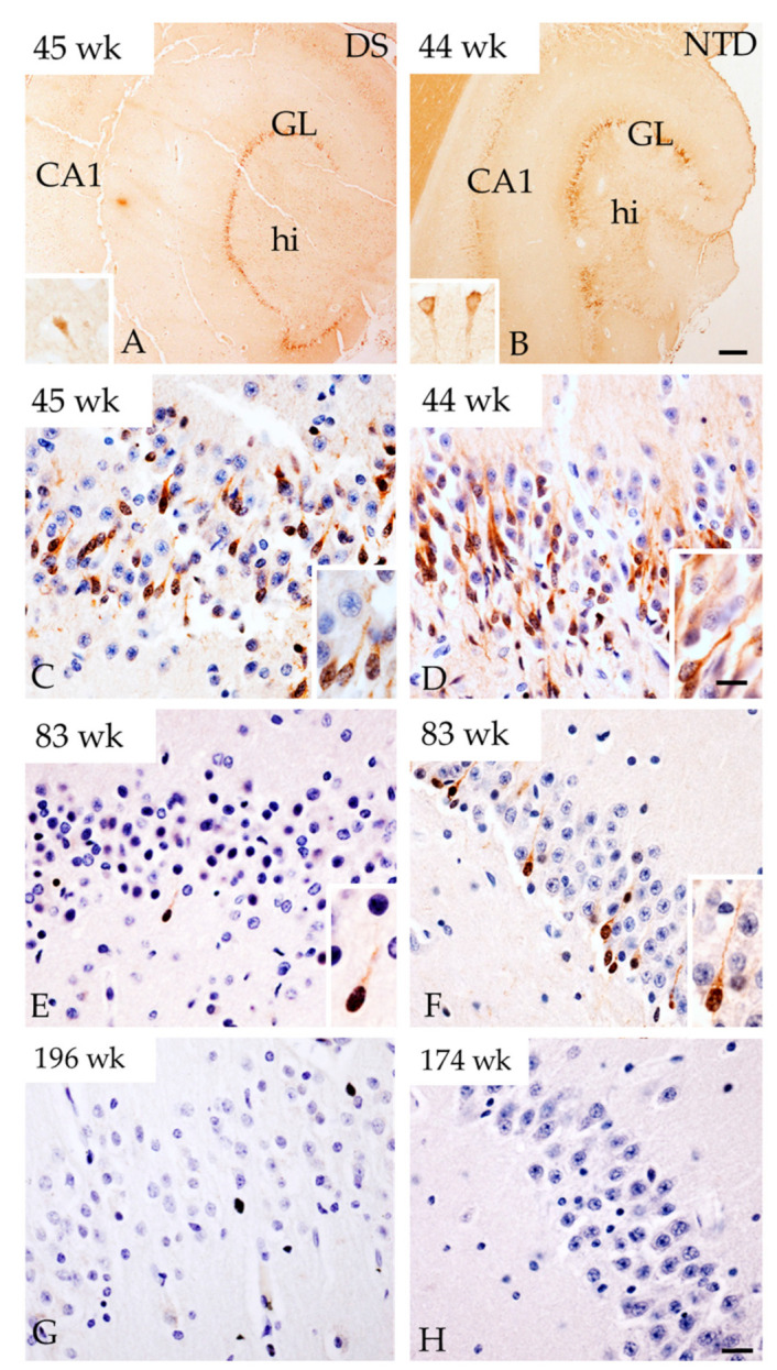
Images showing strong DCX immunoreactivity in the DG GL at week 45 in DS (A) and at 44 weeks in NTD (B). Insets in panel (A) and (B) show DCX-ir CA1 pyramidal cells in a DS 45 and NTD 44 week cases, respectively. Images showing DCX-ir cells in the GL at postnatal weeks 45, 83 and 196 in DS and at 44, 83, 174 weeks in NTD cases. Insets in panels (C–F) show details of DCX positive cells in the GL. Note that less cells were positive for DCX in DS at 45 (C) and 83 (E) postnatal weeks compared to 44 (D) and 83 (F) weeks in NTD and were absent at 196 (G) and 174 (H) weeks in DS and NTD, respectively. Section in (C–H) were counterstained with hematoxylin. Abbreviations: CA1 = hippocampal subfield CA1, GL = granule cell layer, hi = hilus. Scale bar in (B) = 500 µm and applies to (A), and in (H) = 25 µm applies to (C–G) panels, in (D) inset = 10 µm and applies to insets of (A–F).