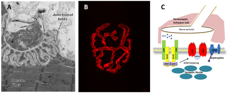
Figure 1.
The neuromuscular junction (NMJ) in skeletal muscle. (A) Representative electron microscopy image showing ultrastructure of the NMJ. (B) Representative confocal microscopy image of fluorescent stain with an acetylcholine receptor binding neurotoxin (α-Bungarotoxin, BTX, red). (C) Illustration describing the components of the NMJ. Perisynaptic Schwann cells are glial regulators of NMJ structure and function. Agrin interacts with lipoprotein receptor-related protein 4 (Lrp4), which activates muscle-specific kinase (MuSK). MuSK, a transmembrane tyrosine kinase, through signaling of Dok-7 and rapsyn, drives acetylcholine receptor (AChR) clustering. Dystrophin and its associated glycoprotein complex (DGC), which is present throughout the muscle fiber membrane, accumulate underneath the post-synaptic membrane.

