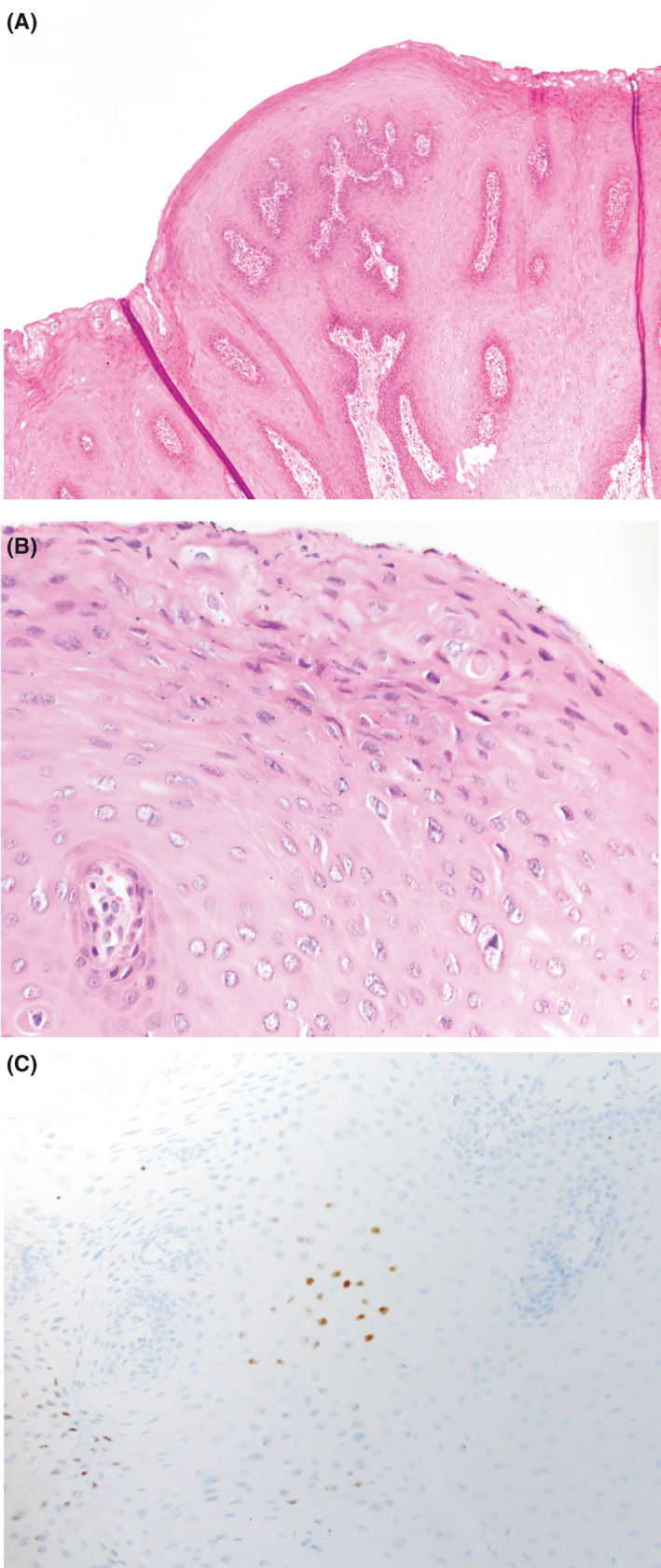
FIGURE 2.

Sections demonstrate a squamous lesion with papillary architecture (A, H&E, 100× magnification). There was focal koilocytic atypia in the lesion (B, H&E, 400× magnification). RNA in situ hybridization showed lesional cells were positive for low‐risk HPV in a nuclear localization (C, in situ hybridization, 400× magnification), as seen in HPV‐mediated proliferations
