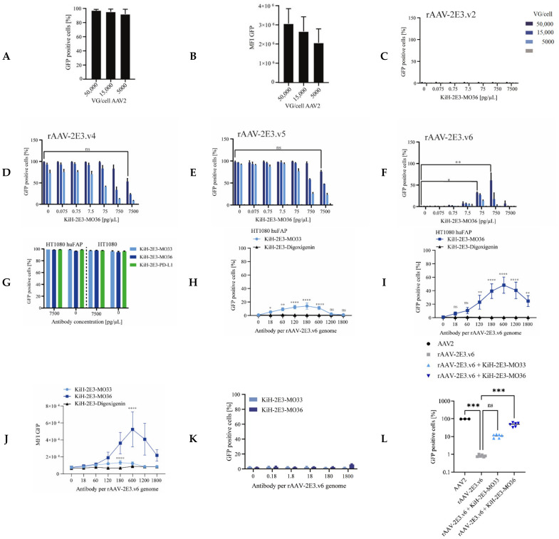
Figure 5.
Retargeting novel rAAV-2E3 to FAP expressing cells using KiH bispecific antibodies. HT1080 huFAP cells were treated with AAV viral variants for 3 days following flow cytometry analysis. (A) AAV2 transduction with titrated VG/cell and analysis of GFP positive proportion and (B) MFI levels. Data show mean + SD. (C) Proportion of cellular GFP expression after retargeting with rAAV-2E3.v2, (D) rAAV-2E3.v4, (E) rAAV-2E3.v5, and (F) rAAV-2E3.v6 complexed with KiH-2E3-MO36. (D–F) share legend shown in (C). Data show mean + SD of two independent experiments, one-way ANOVA, uncorrected Dunn’s test, * p < 0.05; ** p < 0.01, ns= non-significant. (G) Bispecific antibodies pre-incubated with AAV capsids did not affect GFP expression levels of transduced cells. Data show mean + SD. (H) Titration of KiH-2E3-MO33, (I) KiH-2E3-MO36, and isotype control rAAV-2E3.v6 (VG/cell 50,000) and analysis of percentage of GFP expressing cells and (J) MFI levels. Data show mean + SD of three independent experiments, one-way ANOVA, uncorrected Dunn’s test groups compared 0.0 pg/µL KiH-2E3, * p < 0.05; ** p < 0.01; **** p < 0.0001, ns = non-significant. (K) rAAV-2E3.v6 complexed with KiH-2E3-MO33 or KiH-2E3-MO36 and incubation on HT1080 huFAP negative cells did not result in GFP expression. Data show mean + SD. (L) Direct comparison of HT1080 huFAP GFP expression levels after transduction with AAV2, rAAV-2E3.v6, rAAV-2E3.v6:KiH-2E3-MO33, and rAAV-2E3.v6:KiH-2E3-MO36 at 50,000 VG/cell and 2.5 ng/µL bispecific antibody, one-way ANOVA, uncorrected Dunn’s test *** p < 0.001, ns = non-significant.

