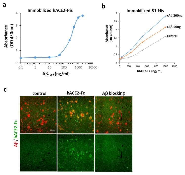
Figure 2.
Interactions between Aβ1-42 and ACE2. (a) Aβ1-42 bound to immobilized hACE2-His (the estimated EC50 is 274–363 ng/mL), suggesting an apparent interaction between Aβ1-42 and hACE2. (b) ACE2-Fc bound to immobilized S1 of SARS-CoV-2 in the absence of Aβ1-42 (control), and this binding was increased by pre-incubation of immobilized S1 of SARS-CoV-2 with Aβ1-42 at 50 and 200 ng, whose binding potency was compared by OD. (c) As shown by confocal microscopy, exogenous hACE2-Fc (green) co-localized with Aβ plaques (red) in the brain tissue sections of APP/PS1 mice (vehicle shown in left panels vs. hACE-Fc in middle panels). Pre-incubation of hACE-Fc with Aβ1-42 (right panel) completely blocked hACE2-Fc/Aβ plaque interaction (as indicated by Aβ blocking). Scale bar: 100 μm. Data of the binding assay are presented as mean values from two independent experiments.

