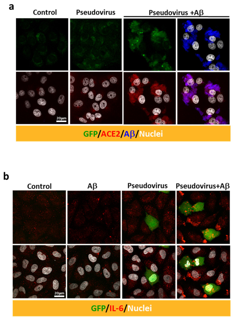
Figure 4.
Aβ1-42 increases the intracellular immuno-reactivity of IL-6 in a SARS-CoV-2 pseudovirus infection model. (a) Immuno-reactivity of endogenous ACE2 (in red) was detected at relatively low levels in the controls. Treatment with Aβ1-42 (10 µg/mL) and SARS-CoV-2 pseudovirus increased the expression of ACE2 (red, lower panel) as well as the co-localization of ACE2 and Aβ1-42 (in blue) in Vero E6 cells. (b) Intracellular IL-6 expression was evaluated by confocal microscopy after infection with SARS-CoV-2 pseudovirus in the presence or the absence of Aβ1-42 (50 µg/mL) in human A549 alveolar epithelial cells. GFP (in green) was abundantly expressed in a few cells 17 h post-infection. Minimal intracellular IL-6 immuno-reactivity was observed in cells receiving either Aβ1-42 or SARS-CoV-2 pseudovirus alone. In contrast, infection with SARS-CoV-2 pseudovirus in the presence of Aβ1-42 increased intracellular IL-6 immuno-reactivity (in red). Scale bars: 20 μm.

