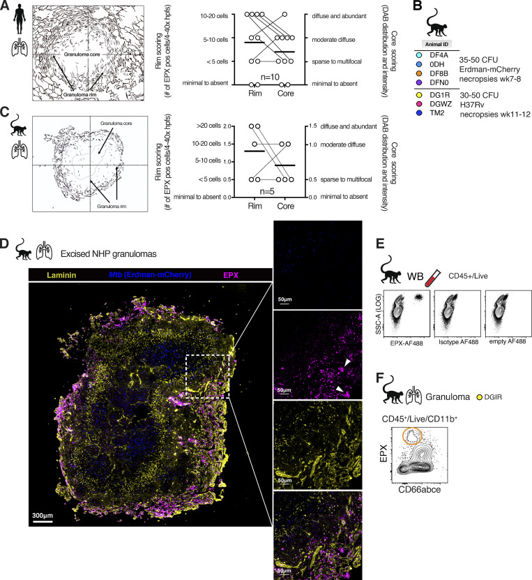
Figure S2.
Human and NHP TB lesions assessed by H&E and EPX immunohistological and flow cytometric staining. (A) Rome cohort: Histological scoring of eosinophil distribution in human TB lesion on blinded specimens (n = 10). (B) Animal IDs and infection dose and strains for NHP Mtb infections (male and female, n = 7, two independent experiments). (C) Granuloma outline and legend for histological scoring of NHP granulomas on blinded specimens (n = 5). (D) EPX immunofluorescence staining on thin sections from paraffin-embedded granulomas after Erdman-mCherry (blue) Mtb infection. (E) Isotype and fluorescence minus one (empty) control of EPX–AF488 staining in NHP WB. (F) Example EPX FACS staining in granulomas from rhesus macaques. DAB, 3,3′-diaminobenzidine; hpfs, high-power fields; SSC, side scatter.

