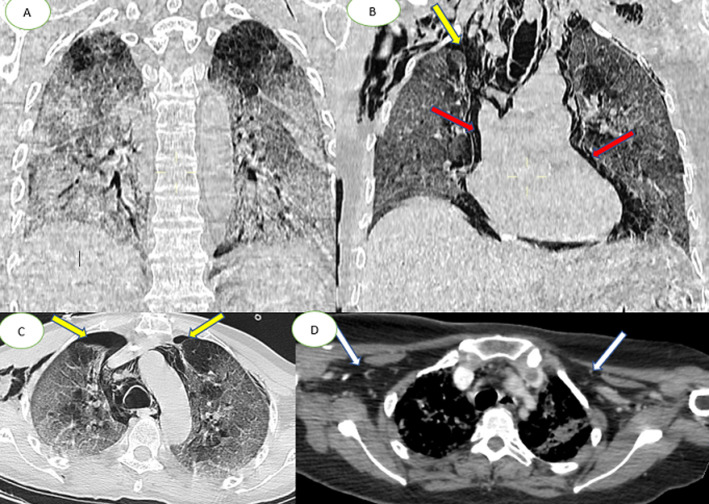
Figure 2.
chest CT scan in coronal (A, B) axial (C, D) and lung parenchymal windows showing subpleural ground-glass opacities associated with multiple areas of consolidation as well as the presence of bilateral pneumothorax, (yellow arrows) pneumomediastinum, (red arrows); extensive bilateral subcutaneous emphysema (white arrows)

