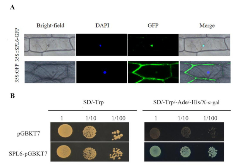
Figure 3.

Subcellular location and transcription activity analysis of SmSPL6 protein. (A) Subcellular location of SmSPL6. The fluorescence signal was observed via laser scanning confocal microscope. DAPI, 4′, 6-diamidino-2-phenylindole is a blue fluorescent DNA stain that was used for indicating nucleus region. (B) Transcription activity analysis of SmSPL6 protein. Yeast colonies with three different dilutions were grown on the SD/-Trp medium, then spotted on the SD/-Trp/-Ade/-His/X-α-gal. X-α-gal: 5-Bromo-4-chloro-3-indolyl-α-D-galactoside medium, the color reaction substrate of α-galactosidase.
