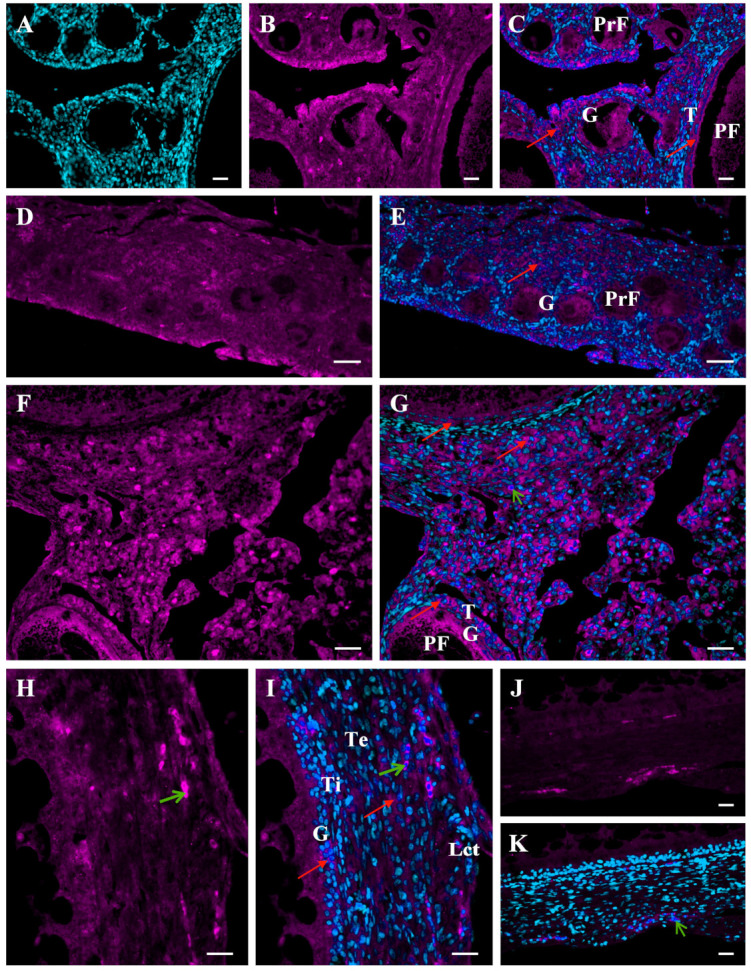
Figure 2.

Immunofluorescent localization of the MMP-2 protein in the ovary of a growing chicken. Representative micrographs of the chicken ovary obtained about 6 weeks (A–C) and 4 weeks (D,E) before sexual maturation and of the ovarian stroma obtained from a mature hen (F,G). The wall of the yellowish follicle obtained from the ovary about two weeks before maturation (H–K). Positive fluorescence for MMP-2 (magenta; red arrows) present in ovarian stromal cells as well as the entire wall of primordial follicles (PrF; composed of one layer of granulosa cells), primary follicles (PF; composed of one layer of granulosa cells and a thin theca layer), and yellowish follicles (composed of several layers of granulosa cells, theca interna and externa, and epithelium with loose connective tissue). Negative control sections incubated without the primary antibody (Abcam, Cambridge, UK) do not exhibit positive staining (J,K), excluding red blood cells which always show nonspecific fluorescence (green arrows). DAPI staining (cyan) of the nucleus only (A). Specific immunoreactivity for MMP-2 (B,D,F,H). Merger of DAPI and MMP-2 (C,E,G,I,K). Abbreviations: G, granulosa; T, theca; Lct, epithelium with loose connective tissue; Ti, theca interna; Te, theca externa. Scale bars = 20 µm.
