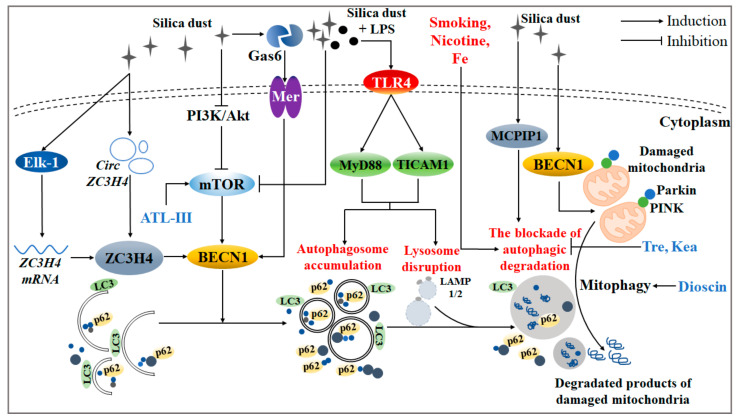
Figure 1.
Related molecular mechanisms and effects during the process of silica-induced autophagy. Silica invasion led to the accumulation of autophagosomes and the disruption of lysosomes, thereby breaking the function of autophagic degradation through the PI3K/Akt/mTOR signaling pathway. Under silica circumstances, ZC3H4 and MCPIP1 both activated autophagy activity. Concretely, ZC3H4 was enhanced not only by the up-regulation of circZC3H4, but also by the transcription of ZC3H4 mRNA via phosphorylation of Elk-1. Furthermore, the interaction of Gas6 and its receptor, Mer, boosted autophagy activity. In addition, Fe atoms were found to be accumulated on the surface of silica, which further magnified the pathological damage of the autophagic process. LPS, nicotine, and habitual smoking all led to the blockade of autophagic degradation. Specifically, TLR4/Myd88 or TLR4/TICAM signaling pathways might be involved in the autophagy induced by silica together with LPS. Particularly, natural products like tre and kea had a protective function in the degradation process of autophagic substrates. When silica invaded, dioscin removed redundant damaged mitochondria through AM mitophagy, and ATL-III protected AM autophagic degradation via an mTOR-dependent signaling pathway.

