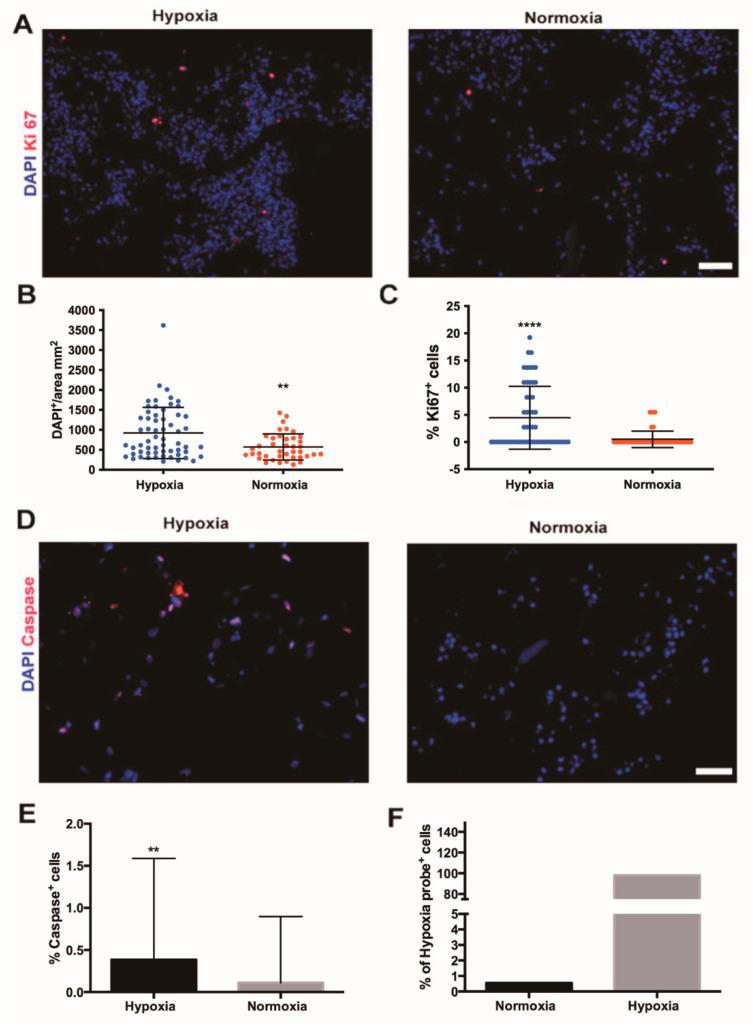
Figure 2.
Effects of perfusion culture under hypoxia or normoxia on human stromal vascular fraction cell proliferation and apoptosis. (A) Immunofluorescence staining for Ki67, the marker of proliferation (red) of perfused SVF cell-based constructs in either hypoxia or in vitro normoxia. Cell nuclei were stained with DAPI (blue). Scale bar = 70 µm. Quantification of total number of SVF cells (DAPI-positive cells) (B) and percentage of proliferating (Ki67-positive) cells (C) in constructs cultured in perfusion culture under high or low oxygen tension. **** p < 0.00001; ** p < 0.001. n = 72 (6 donors). (D) Immunofluorescence staining for marker of apoptosis (cleaved caspase-3, red) of SVF cell-based constructs cultured in hypoxia and in vitro normoxia. Cell nuclei were stained with DAPI (blue). Scale bar = 35 µm. (E) Percentage of apoptotic cells, positive for cleaved caspase-3 in constructs cultured in perfusion culture under high or low oxygen tension. ** p < 0.001. n = 36 (3 donors). (F) Flow cytometry analysis of SVF cells cultured in perfusion in normoxia or hypoxia positive to the pimonidazole. Donor n = 1. Comparison was performed with non-parametric test (Mann–Whitney-U).

