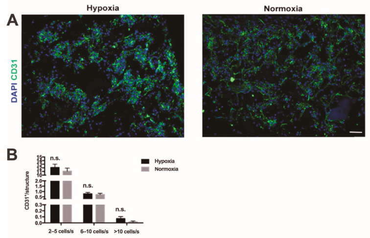
Figure 3.
In vitro endothelial cell organization. (A) Immunofluorescence staining endothelial cell marker (CD31, green) of constructs with SVF cells cultured in hypoxia and in normoxic culture. Cell nuclei were stained with DAPI (blue). Scale bar = 70 µm. (B) Quantification of number of organized endothelial cells in small (3–5 cells per structure), middle-size (5–10 cells per structure), and complex (over 10 cells per structure) cord-like structures. n.s. = not statistically significant; s: structure; n = 72 (6 donors). Comparison was performed with non-parametric test (Mann–Whitney-U).

