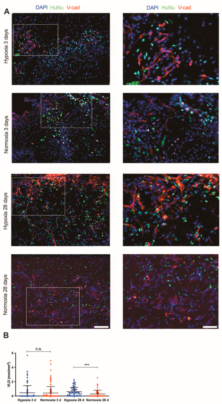
Figure 5.
In vivo vascularization potential of SVF-based patches. (A) Immunofluorescence staining endothelial cells (Ve-caderin, red), human nuclei specific marker (HuNu, green) of constructs generated by SVF cells in severe hypoxia and normoxia perfusion-based culture with investigation time points 3 and 28 days (d) at low (20×) and high (40×) magnifications (scale bars = 50 µm and 25 µm, respectively). (B) Quantification of vessel length density (VLD) of constructs cultured under severe hypoxia or in vitro normoxia after implantation in vivo with investigation time points 3 and 28 days. *** p < 0.001; n.s. = not statistically significant. Comparison was performed with one-way ANOVA. Number of donors: 3 donors (at least duplicate biological samples per donor).

