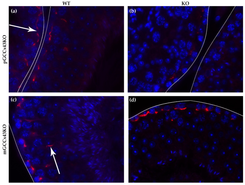
Figure 7.
Representative localization of Connexin43 (Cx43) in adult p- and mGCCx43KO and WT mice. For easier orientation, the basal lamina of the tubules is marked by dotted lines. Cx43 forms a fine linear staining pattern (red) in the basal area of the seminiferous tubules (arrow in (a)) in WT mice of both mouse lines (a,c) resulting from synthesis by both Sertoli cells and basally located germ cells. Furthermore, Cx43 can also be found more apically in the seminiferous epithelium (arrow in (c)) in WT mice. In the KO animals of the pGCCx43KO mouse line (b), the described staining pattern in the basal area of the seminiferous epithelium is also visible; however, the staining intensity seems to be less intense, probably resulting from the lack of Cx43 in the basally located germ cells due to the KO in spermatogonia/early spermatocytes. In the mGCCx43KO mouse line (d), the apical localization of Cx43 could not be detected indicating that the more apically localized germ cells (spermatids) do not synthesize Cx43 following its KO in those cell types. Magnification: 630×.

