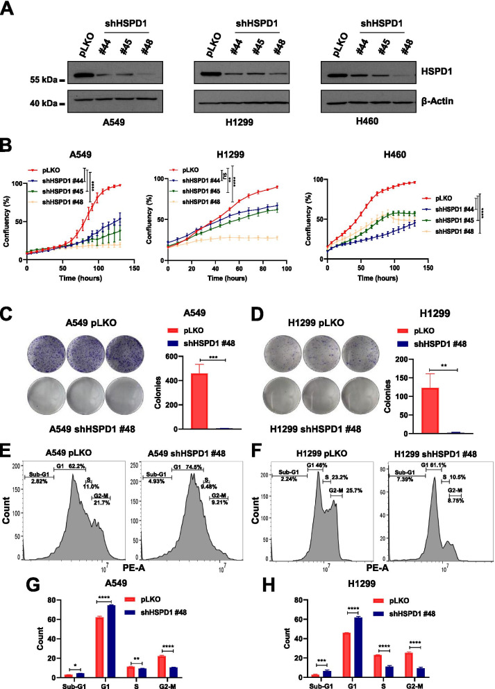
Fig. 2.
HSPD1 knockdown blocks cell proliferation and clonogenic ability of NSCLC cells. A) Western blot analysis of HSPD1 protein levels in A549, H1299 and H460 cells upon infection with 3 independent shRNAs (#44, #45 and #48) targeting HSPD1 compared to scramble-infected cells (pLKO). β-Actin was used as loading control. B) Real-time proliferation curves of A549, H1299 and H460 infected with non-targeting pLKO or shHSPD1. The cells’ confluency plotted over time is shown. P-values are from two-way ANOVA. Points are averages of biological replicates ± SD. ** < 0.01, **** < 0.0001. Colony formation of A549 (C) and H1299 (D) cells with pLKO or shHSPD1, stained with crystal-violet and quantified in triplicates. Bars are average of biological replicates ± SD. P-values are from unpaired t-test. ** < 0.01, *** < 0.001. FACS plots of A549 (E) and H1299 (F) cells infected with pLKO or shHSPD1 and stained with PI for cell cycle analysis. Bar graph showing the % of cells in each cell cycle phase of A549 (G) and H1299 (H) cells upon infection with pLKO or shHSPD1. P-values are from two-way ANOVA. Bars are average of biological replicates ± SD. * < 0.05, ** < 0.01, *** < 0.001, **** < 0.0001

