Fig. 5.
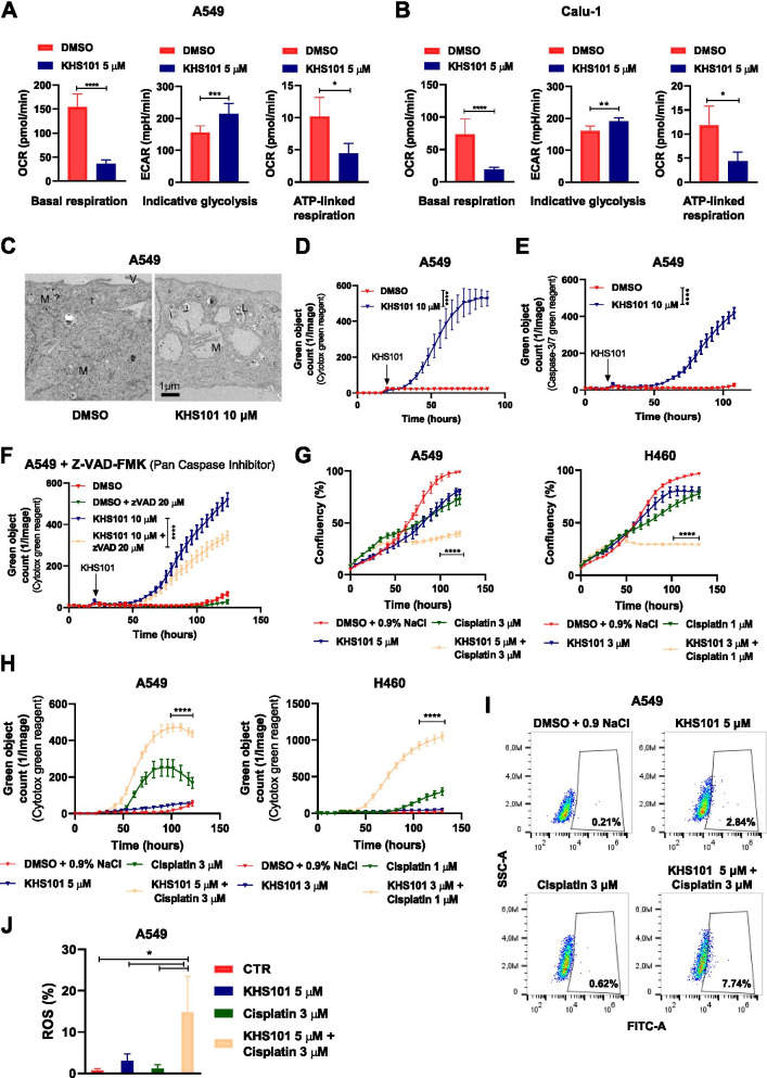
KHS101 induces a metabolic impairment and death in NSCLC cells. Bar graphs showing quantification of basal respiration, indicative glycolysis and ATP-linked respiration in A549 (A) or Calu-1 (B) treated with KHS101 for 24 h compared to control cells (DMSO). Bars are average values of biological replicates ± SD. P-values are from unpaired t-test. * < 0.05, ** < 0.01, *** < 0.001, **** < 0.0001. C) Electron microscopy images of A549 treated 96 h either with vehicle (DMSO) or KHS101 10 μM (M: mitochondria, V: microvilli). Dead cell quantification as green object count using Cytotox green reagent (D) or caspse-3/7 reagent (E) of vehicle or KHS101 treated A549 cells. F) Dead cell quantification as green object count of A549 treated either with DMSO or KHS101 in combination with pan-caspase inhibitor Z-VAD-FMK. In D, E and F points are average of biological replicates ± SD. P-value are from two-way ANOVA. **** < 0.0001. G) Real-time proliferation curves of A549 and H460 treated with KHS101 and/or cisplatin compared to control cells. Cell death is shown as green object count in H). Points are average values of biological replicates ± SD. P-values are from two-way ANOVA and analyzed comparing the combination of drugs to control and the respective drug alone. **** < 0.0001. Percentage of ROS-positive cells in A549 cells treated with KHS101 and/or cisplatin for 24 h are shown in FACS plots (I) and a graph bar (J). P-values are from one-way ANOVA. Bars are average of biological replicates ± SD. * < 0.05

