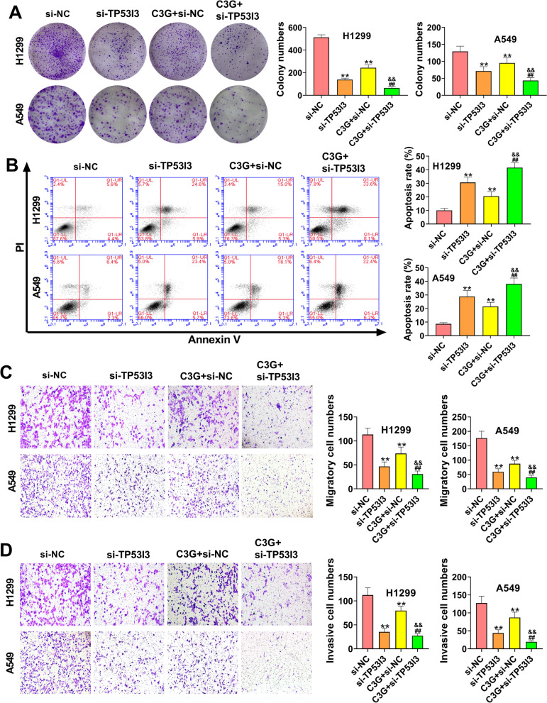
Fig. 5.
C3G inhibited the proliferation, migration, and invasion, and promoted apoptosis in H1299 and A549 cells by downregulating TP53I3. A After the treatment of C3G (40 μM) or si-TP53I3, the cell clones of H1299 and A549 cells were determined by colony formation assay. B After the treatment of C3G (40 μM) or si-TP53I3, the apoptosis of H1299 and A549 cells was detected by flow cytometry. C After the treatment of C3G (40 μM) or si-TP53I3, the migration of H1299 and A549 cells was detected by transwell assay. D After the treatment of C3G (40 μM) or si-TP53I3, the invasion of H1299 and A549 cells was detected by transwell assay. **P < 0.01, vs. si-NC group; ##P < 0.01, vs. si-TP53I3 group; &&P < 0.01, vs. C3G + si-NC group

