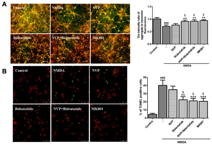
Figure 4.
Involvement of NR2B-containing NMDA receptors in anti-apoptosis effects of bidentatide: (A) representative fluorescence micrographs of JC-1 staining for (aggregate in red, monomer in green, and the merge in yellow) and bar graph of the fluorescence intensity ratio of aggregate-to-monomer in primary cultured hippocampal neurons upon no treatment, exposure to NMDA alone, pretreatment with NVP-AAM077 (100 nM) for 30 min and then exposure to NMDA, pretreatment with bidentatide (100 nM) for 30 min and then exposure to NMDA, pretreatment with NVP-AAM077 (100 nM) and bidentatide (100 nM) for 30 min and then exposure to NMDA, and pretreatment with 10 μM MK801 for 30 min and then exposure to NMDA; scale bar, 100 μm; (B) representative fluorescence micrographs of TUNEL staining (PI staining in red, TUNEL-positive staining in green, the merged area in yellow) and bar graph of the percentage of TUNEL-positive cells in primary cultured hippocampal neurons upon different treatments as mentioned above. Yellow indicates merged PI staining (red) and TUNEL-positive staining (green); scale bar, 75 μm; ### p < 0.001 verses control; ** p < 0.01, and *** p < 0.001 verses NMDA alone; $ p < 0.05 versus pretreatment with NVP-AAM077 plus NMDA.

