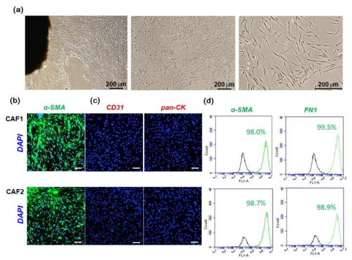
Figure 1.
Immunocytochemical analysis of fibroblasts cultured from HNSCC patients. The stroma adjacent to the tumor was cut into the smallest possible pieces in sterile DMEM and seeded in 10-cm tissue culture dishes supplemented with 10% FBS and then cultured for 2–3 weeks. (a) The representative image was taken by phase-contrast microscope. (b,c) Cells were seeded in 6-well plates containing cover slides and immunostained with antibodies specific for fibroblasts (α-SMA), endothelial cells (CD-31), and epithelial cells (pan-cytokeratin). The white scale bars represent 100 μm. (d) α-SMA-positive or fibronectin (FN1)-positive fibroblasts were analyzed with a flow cytometer. The experiments were performed three times and representative images are shown.

