Figure 6.
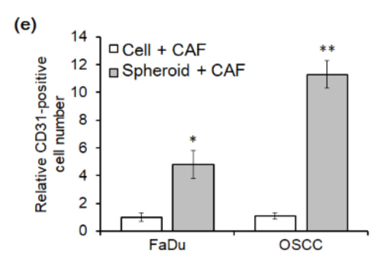
Immunohistochemical analysis of mice tumor tissues stained with anti-CD31 antibody, a specific endothelial marker (a–d). Representative CD31-positive cells were indicated with black arrows. (e) The CD31-positive cells were counted in immunostained mice tumor tissues (* p < 0.05, ** p < 0.01).


