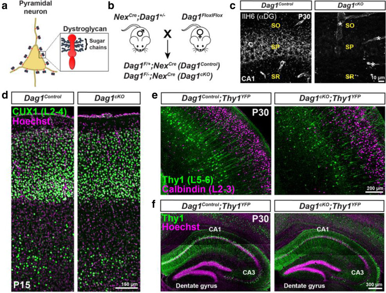
Fig. 1.
Neuronal Dystroglycan is not required for pyramidal neuron migration. (a) Schematic of Dystroglycan on pyramidal neurons. Inset shows the structure of Dystroglycan and sugar chain moieties present on the extracellular subunit. (b) Mouse breeding scheme for generating pyramidal neuron-specific Dag1 conditional knockout mice using NexCre driver mice. (c) Immunostaining for Dystroglycan in the hippocampal CA1 region of P30 Dag1Control mice (left panel) shows punctate Dystroglycan protein on the soma and proximal dendrites of pyramidal neurons, whereas Dag1cKO mice (right panel) lack perisomatic staining. Asterisks denote Dystroglycan staining on blood vessels which is retained in Dag1cKO mice. (d) Coronal sections from P15 Dag1Control and Dag1cKO cortex were immunostained for upper layer marker CUX1 (L2–4). (e) Coronal sections of the cortex from P30 Dag1Control and Dag1cKO mice crossed with a Thy1YFP reporter mouse to sparsely label layer 5–6 pyramidal neurons (green) and stained for Calbindin (magenta) to label layer 2–3 pyramidal neurons. (f) Coronal sections of the hippocampus from P30 Dag1Control and Dag1cKO mice crossed with a Thy1YFP reporter mouse to label excitatory neurons (green) in the CA regions and dentate gyrus

