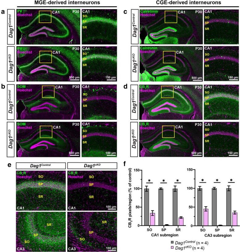
Fig. 2.
CCK+ interneurons are selectively reduced in mice lacking Dystroglycan from pyramidal neurons. (a-b) Immunostaining for medial ganglionic eminence (MGE)-derived interneuron markers (green) parvalbumin (PV) (a) and somatostatin (SOM) (b) show normal innervation of the hippocampus in P30 Dag1Control and Dag1cKO mice. Insets (yellow boxed regions) show enlarged images of the CA1. (c-d) Immunostaining for caudal ganglionic eminence (CGE)-derived interneuron markers (green) Calretinin (c), and CB1R (d) show normal innervation of Calretinin interneurons in Dag1Control and Dag1cKO mice, whereas CB1R is largely absent from the CA regions of Dag1cKO mice. Insets (yellow boxed regions) show enlarged images of the CA1. (e) Immunostaining for CB1R in hippocampal CA1 (top) and CA3 (bottom) of P30 Dag1Control and Dag1cKO mice. (f) Quantification of CB1R pixels for each CA layer of the CA1 and CA3 shows a significant reduction in CB1R staining in Dag1cKO mice (*P < 0.05, unpaired two-tailed Student’s t-test; n = 4 mice/genotype). Data are presented as mean values ± s.e.m. Data are normalized to Dag1Control signal in each CA layer. CA layers: SO, stratum oriens; SP, stratum pyramidale; SR, stratum radiatum

