Fig. 3.
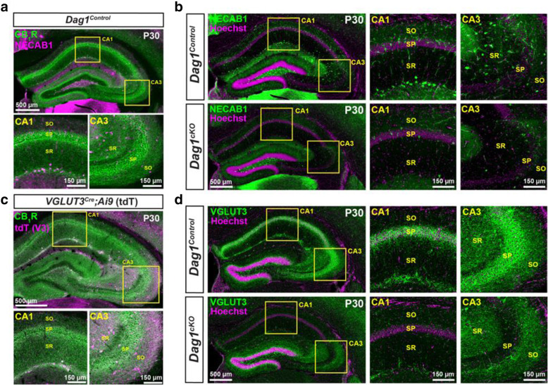
Cell body and synaptic markers for CCK+ interneurons are reduced in Dag1cKO mice. (a) Immunostaining showing the co-localization of CB1R (green) and NECAB1 (magenta) in CCK+ interneurons. Insets (yellow boxed regions) show enlarged images of the CA1 and CA3. (b) Immunostaining for NECAB1 (green) shows a reduction of NECAB1+ interneurons in the hippocampus of P30 Dag1cKO mice. Insets (yellow boxed regions) show enlarged images of the CA1 and CA3. (c) Immunostaining of hippocampal sections from VGLUT3Cre mice crossed with a Lox-STOP-Lox-tdTomato (Ai9) reporter mouse showing the co-localization of CB1R (green) and VGLUT3 (magenta) in a subset of CCK+ interneurons. Insets (yellow boxed regions) show enlarged images of the CA1 and CA3. (d) Immunostaining for VGLUT3 (green) shows a reduction of CCK+ interneuron synaptic terminals in the hippocampus of P30 Dag1cKO mice. Insets (yellow boxed regions) show enlarged images of the CA1 and CA3. CA layers: SO, stratum oriens; SP, stratum pyramidale; SR, stratum radiatum

