
Figure 3. Halicin is a broad-spectrum bactericidal antibiotic.
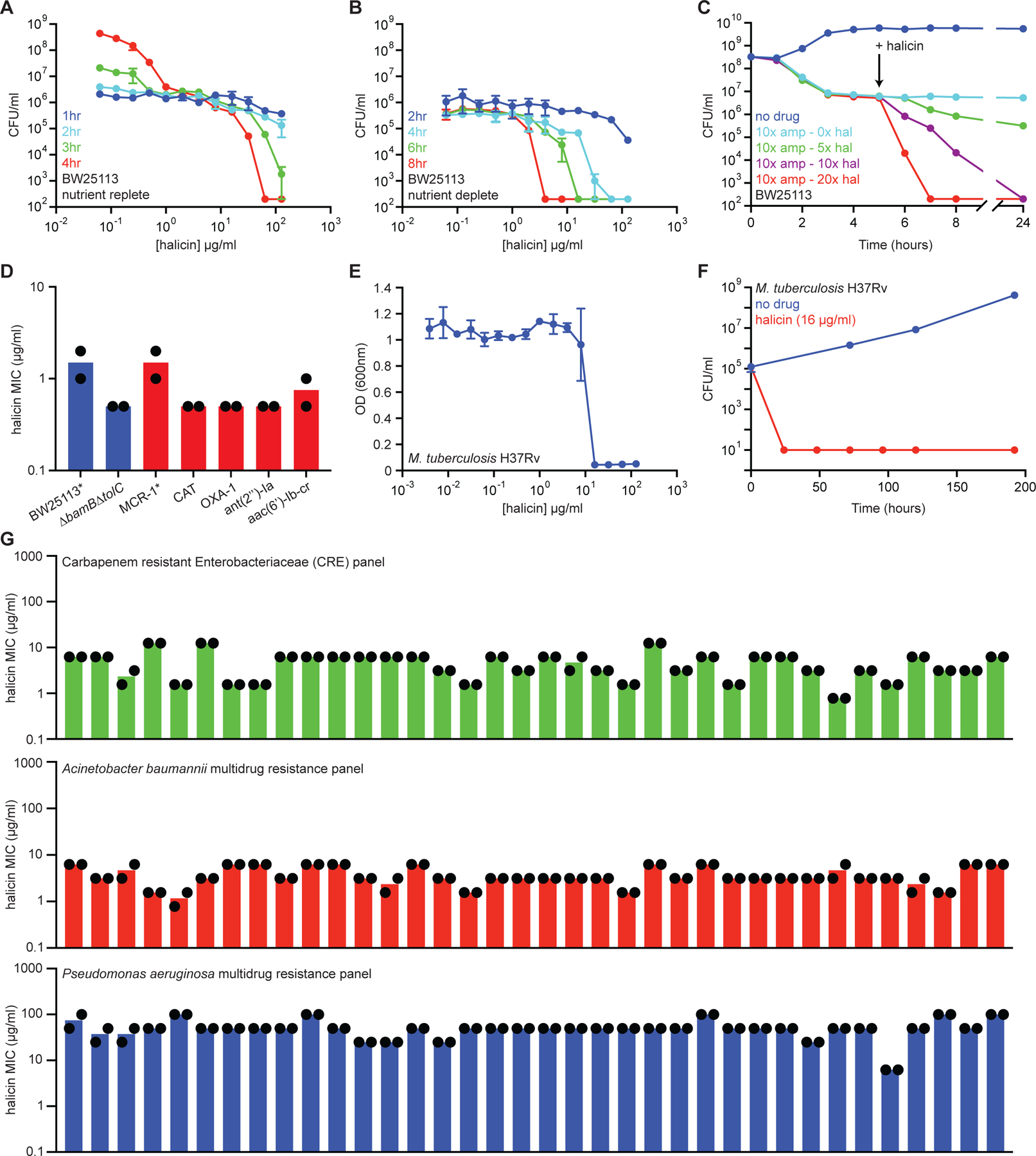
(A) Killing of E. coli in LB media in the presence of varying concentrations of halicin after 1 hr (blue), 2 hr (cyan), 3 hr (green), and 4 hr (red). The initial cell density is ~106 CFU/ml. Shown is the mean of two biological replicates. Bars denote absolute error. (B) Killing of E. coli in PBS in the presence of varying concentrations of halicin after 2 hr (blue), 4 hr (cyan), 6 hr (green), and 8 hr (red). The initial cell density is ~106 CFU/ml. Shown is the mean of two biological replicates. Bars denote absolute error. (C) Killing of E. coli persisters by halicin after treatment with 10 µg/ml (10x MIC) of ampicillin. Light blue is no halicin. Green is 5x MIC halicin. Blue is 10x MIC halicin. Red is 20x MIC halicin. Shown is the mean of two biological replicates. Bars denote absolute error. (D) MIC of halicin against E. coli strains harboring a range of antibiotic-resistance determinants. The mcr-1 gene was expressed in E. coli BW25113. All other resistance genes were expressed in E. coli BW25113 ∆bamB∆tolC. Experiments were conducted with two biological replicates. (E) Growth inhibition of M. tuberculosis by halicin. Shown is the mean of three biological replicates. Bars denote standard deviation. (F) Killing of M. tuberculosis by halicin in 7H9 media at 16 µg/ml (1x MIC). Shown is the mean of three biological replicates. Bars denote standard deviation. (G) MIC of halicin against 36-strain panels of CRE isolates (green), A. baumannii isolates (red), and P. aeruginosa isolates (blue). Experiments were conducted with two biological replicates. See also Figure S2, Table S3.