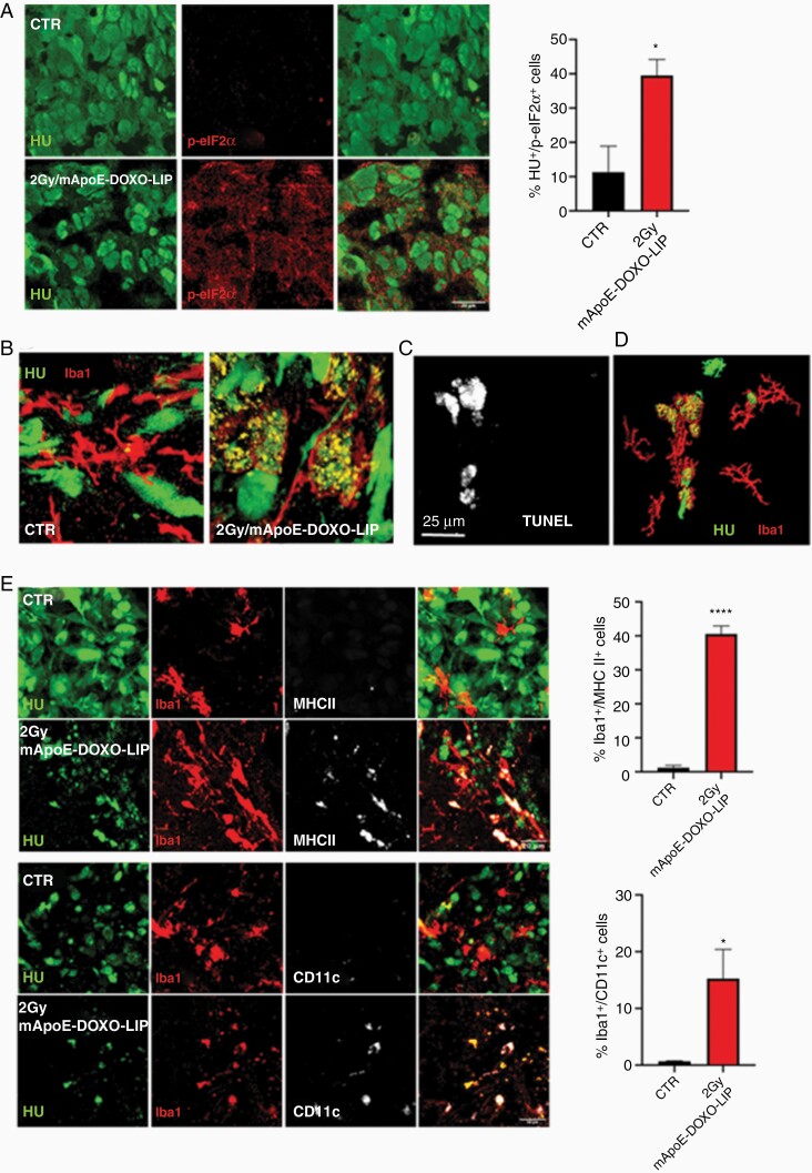
Figure 6.
GSC cell death and immune activation. (A) Detection and quantification of the ICD marker phospho-eIF2α (p-eIF2α) in GSC1tumors, Human Nuclei (HU) positive cells, from untreated (CTR) and 2Gy/mApoE-DOXO-LIP treated mice. p-eIF2α quantification was carried out on 3 brain coronal images each condition acquired using a DMi8 fluorescent microscope and a Leica Application Suite X (LAS X) imaging system (Leica Microsystems). (B) Representative confocal images of Iba-positive, tumor infiltrating microglia/macrophage in untreated (CTR) and 2Gy/mApoE-DOXO-LIP treated mice. (C and D) In situ TUNEL detection within Iba1-positive cells in GSC tumor border and 3D rendering of panel C; (E) Representative confocal images and quantification of the dendritic markers MHCII and CD11c in Iba1-positive cells in untreated (CTR) and 2Gy/mApoE-DOXO-LIP treated mice. Quantification has been performed as in panel A. *P < .05, ****P < .0001.

