Figure 3.

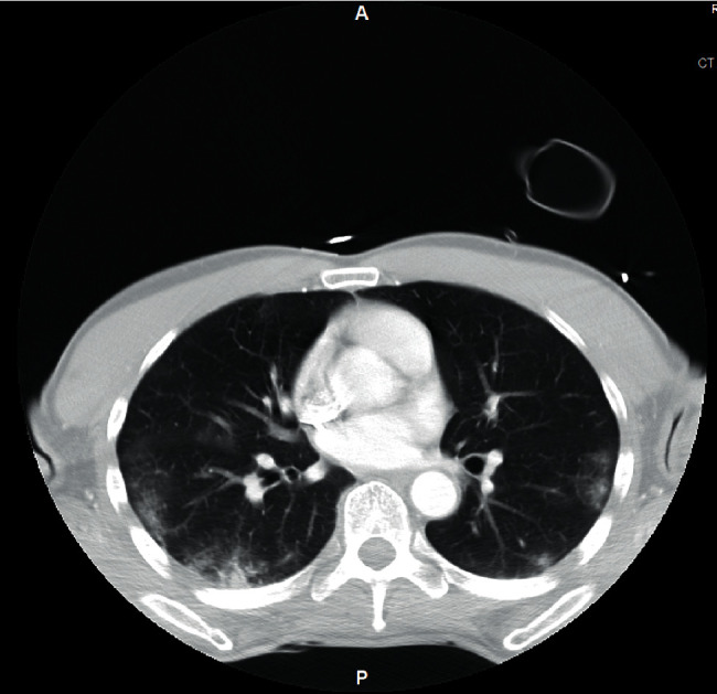
Chest CT of the COVID-19 patient showing patchy and peripheral ground glass opacities mid to lower lobe predominant (taken from another COVID-19-positive patient for comparison).

Chest CT of the COVID-19 patient showing patchy and peripheral ground glass opacities mid to lower lobe predominant (taken from another COVID-19-positive patient for comparison).