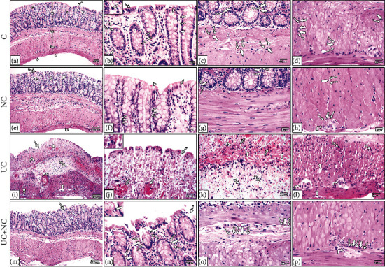
Figure 3.

Photomicrographs of H&E-stained sections in the colon. (a–d) The control group and (e-h) NC group both show the normal structure of the colon; the mucosa (M), submucosa (SM), musculosa (Ms), and serosa (double arrowheads). The mucosa has regularly arranged closely backed tubular crypts (C) the crypts lines by simple columnar absorptive cells (arrow) with numerous goblet cells (arrowhead). The surface shows intact surface epithelium, and the glands are resting on a thin layer of muscularis mucosa. A thin layer of lamina propria containing numerous lymphocytes is present between crypts. (i–l) The ulcerative colitis group reveals loss of normal mucosal architecture, disturbed crypts (star), loss of surface epithelium and goblet cells (arrow), dilated congested blood vessels (lined arrow), and areas of mucosal ulceration (arrow) with thickening of the underling muscularis mucosa (curved arrows). The submucosa is wide with thick dilated blood vessels (BV). Inflammatory cell infiltration (IFC) is obvious in the mucosa and submucosa. The musculosa reveals separation between muscle bundle (tailed arrow), inflammatory cell infiltration (crossed arrow), and myenteric plexus (right-angled arrow). (m–p) The UC+NC group shows the normal architecture of the colon apart from narrow areas of interrupted surface epithelium (arrow). Note: telocytes appear as spindle shape cells (thick arrow) with small flat nuclei and long telopodes (wavy arrow). (H&E, (a, e, i, and m) X100, (b, c, d, f, g, h, j, k, l, n, o, and p) X400 insert in b, j, and n X1000).
