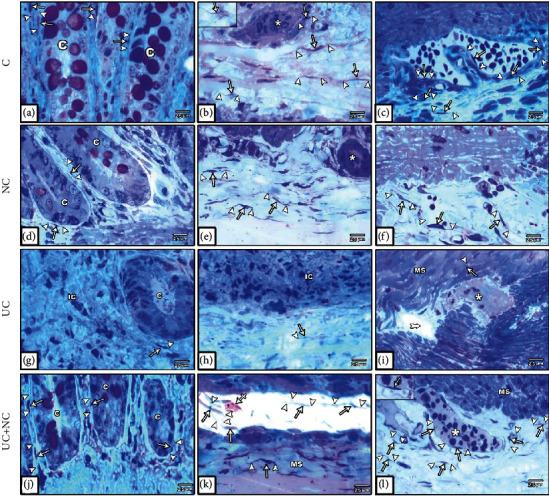
Figure 9.

Photomicrographs of toluidine blue-stained semithin sections in the colon. (a–c) The control group and (d–f) NC group both show numerous TCs (arrow) with long Tps (arrowhead) around the intestinal crypts (C), in the submucosa, musculosa, and myenteric plexus (star). (g–i) The UC group reveals hardly seen TCs with short Tps round the remnant of crypts (C) and completely lost in inflamed areas (IC). Few TCs are hardly seen in the submucosa, the musculosa (MS), and myenteric plexus (star). (j–l) The UC+NC group shows numerous TCs with long Tps around the crypts (C) in the submucosa in contact with Mast cell (crossed arrow), the musculosa (MS), and myenteric plexus (star), (toluidine blue X400 insert in b and i X1000).
