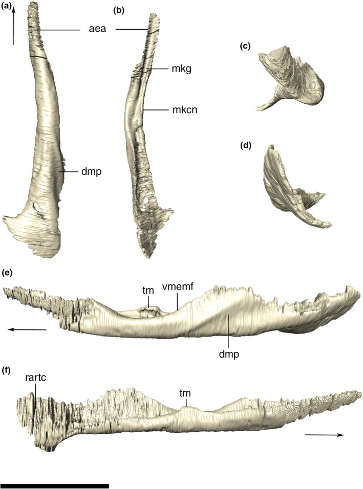
FIGURE 42.

Digitally reconstructed left angular of Aphaurosuchus escharafacies in (a) ventral, (b) dorsal, (c) anterior, (d) posterior, (e) lateral, and (f) medial views. Arrows indicate anterior direction. aea, anterior expansion of the angular; dmp, depression for the insertion of M. pterygoideous; mkcn, Meckelian canal; mkg, Meckelian groove; rartc, retroarticular contact; tm, torose margin; vmemf, ventral margin of the external mandibular fenestra. Scale bar: 10 cm
