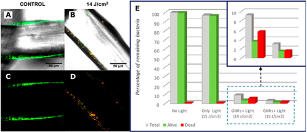
Figure 10. Photothermal Inactivation of S. aureus Biofilm on Surgical Meshes by Au NRs.
(A-D) Fluorescence confocal microscope images at the mesh surfaces for no illumination control (A, C) and group treated with 300 ms-pulsed NIR laser with fluence of 14 J/cm2 (B, D). A, B are a merge of bright field images with fluorescence images, and C, D are pure fluorescence maps. (E) Proportion of alive bacteria at the mesh surface after treatment with different laser fluences.
(A-E) Adapted with permission from Ref. 67. Copyright (2019) American Chemical Society.

